

Beyond Cellular Uptake: How Endosomal Escape Peptides Could Unlock Nucleic Acid Delivery
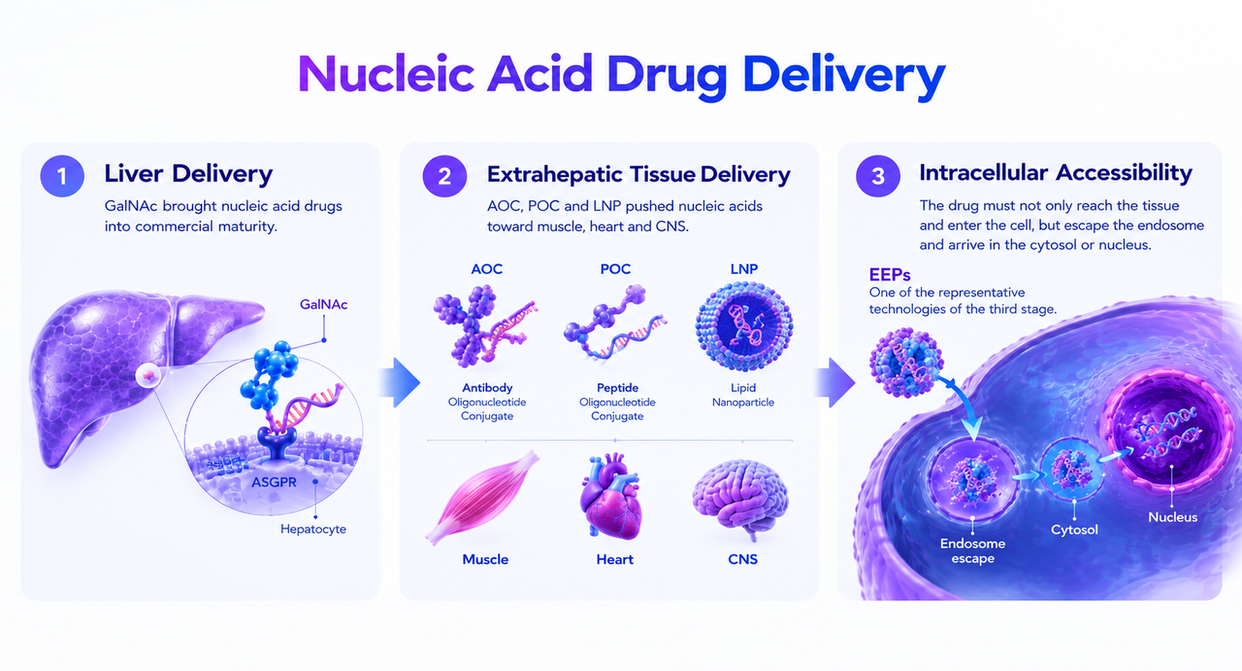
The bottleneck in nucleic acid delivery isn't cell entry. It's getting out of the endosome. Across GalNAc, LNP, AOC and POC, payloads often reach the cell but remain trapped inside endosomes. For siRNA, ASO, PMO and mRNA, no escape means no pharmacology.
Peptides are highly engineerable tools for endosomal escape. Charge, hydrophobicity, pH response, cyclisation and non-natural amino acids provide multiple design levers. A well-designed EEP can combine cellular uptake, membrane disruption, and cytosolic release in one module.
EEPs have moved from academic concept into clinical validation. PepGen and Entrada Therapeutics are leading this transition with their EDO and EEV™ platforms, with two distinct design strategies: engineered linear CPPs and cyclic CPPs, respectively. Programs in DMD and DM1 are now testing the key question: can better endosomal escape translate into meaningful clinical benefit?
EEPs are not substitutes for AOC, LNP or GalNAc; they can become the enabling release layer within these platforms. Reaching the tissue is necessary. Achieving broad cellular coverage, efficient endosomal escape and productive cytosolic delivery is what drives pharmacology. The winning platforms will be those that convert targeting into functional intracellular exposure — at a clinically viable safety margin.
1. Getting into the Cell is not Enough: The Endosomal Bottleneck in Nucleic Acid Delivery
For several years, the delivery story in nucleic acid therapeutics has revolved around tissue targeting. GalNAc showed that hepatocyte delivery could be highly engineered; LNPs give mRNA and siRNA a realistic basis for systemic administration; AOC and POC pushed the boundary further into muscle, heart and the central nervous system. Industrial conversation has correspondingly focused on reaching the target organ, binding the target receptor, entering the target cell.
But for a nucleic acid drug the true endpoint of delivery is not the plasma membrane. It is the cytosol, the nucleus, or a specific subcellular complex. siRNA must reach the cytosol and load into RISC before it can direct degradation of the target mRNA. An ASO or PMO acting on splicing must go further and reach the nucleus to engage pre-mRNA. An mRNA drug must be released into the cytosol to be translated by ribosomes. Entering the cell is not enough; the payload must enter the correct intracellular space.
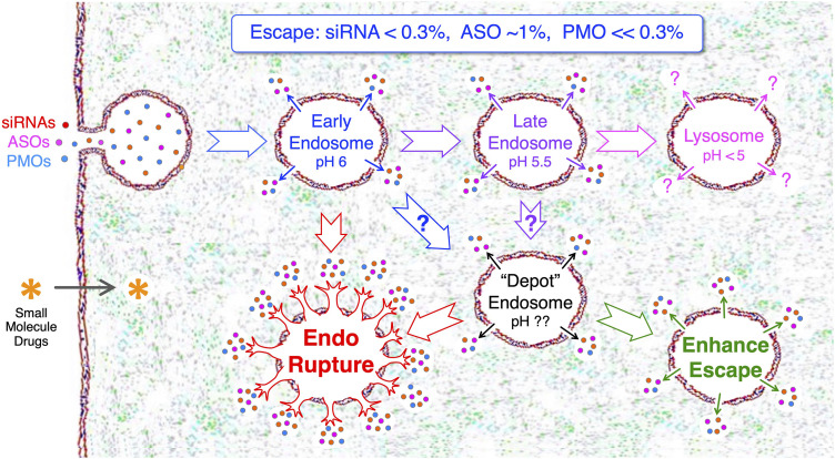
Most nucleic acid drugs enter cells through endocytic routes. Whether by ASGPR-mediated uptake after GalNAc binding, TfR1-mediated delivery to muscle or across the BBB, or CPP- and LNP-driven uptake, the drug's first intracellular destination is usually an early endosome rather than the cytosol. Early endosomes then acidify, mature into late endosomes, and fuse with lysosomes. A payload that does not escape during this window is confined to the endosomal lumen and routed into degradation, the so-called endosomal trap.
Of the nucleic acid drug that enters cells by endocytosis, only around 1% is successfully released into the cytosol. Even a delivery system that looks excellent in cellular uptake assays does not necessarily produce strong pharmacology. Fluorescence showing that a molecule has entered the cell and evidence that it has reached the cytosol or nucleus are two entirely different questions.
This is why so many delivery systems lose potency moving from in vitro to in vivo, and from mouse to human. Uptake can increase by adding positive charge, raising lipophilicity or strengthening receptor binding — but each of these also tends to increase endosomal retention, lysosomal degradation and non-specific toxicity. The difficult part is releasing the drug from the endosome without sacrificing safety.
This is especially visible in muscle disease. DMD and DM1 are not focal lesions; the target tissue is the whole of skeletal muscle, the myocardium, and in some respects multiple systems. A drug that reaches muscle interstitium but cannot efficiently enter myofibers and their nuclei will not generate sufficient splice correction or protein restoration. The limited efficacy of conventional naked PMO exon-skipping drugs is in large part attributable to insufficient muscle uptake and endosomal escape.
Endosomal escape is the true last mile of nucleic acid delivery.
2. From CPPs to EEPs: How Peptide Delivery is Evolving
Endosomal escape peptides did not appear out of nowhere. Their predecessors are cell-penetrating peptides (CPPs) that have been studied for decades. The first generation, HIV TAT peptide, penetratin, R8/R9, were typically rich in arginine and lysine, interacting with anionic components of the cell surface such as glycosaminoglycans and phospholipid head groups to promote uptake. Their emergence established that a short peptide could act as a delivery module and carry otherwise membrane-impermeable macromolecules into cells.
The limitation surfaced quickly: entering a cell is not the same as reaching the cytosol. Many CPP-cargo complexes were taken up abundantly yet remained in endosomes and lysosomes. Increasing positive charge improved uptake but also increased non-specific binding, plasma protein adsorption, renal exposure and cytotoxicity — an unattractive trade for a drug intended for chronic repeat dosing.
Design logic therefore shifted from membrane penetration toward endosomal escape. Second-generation strategies introduced additional functional modules: histidines to add buffering capacity in the acidifying endosome; hydrophobic residues to strengthen membrane insertion; lipidation to raise membrane affinity; pH-responsive architecture so that the peptide remains relatively inert at neutral pH and becomes membrane-active only after endosomal acidification.
Platforms that reached the clinic engineered further. The peptide is no longer a simple cationic sequence but a medicinal-chemistry-optimized delivery unit that must be stable in plasma, distribute in tissue, be taken up by cells, escape the endosome, release in the cytosol or nucleus, and remain manufacturable with a safety window compatible with repeat dosing.
Conceptually, the EEP is an upgraded CPP: where CPPs emphasize getting into the cell, EEPs emphasize getting out of the endosome. This matters especially for oligonucleotides, which are charged, hydrophilic, and conformationally rigid, and cannot cross a lipid bilayer freely. Without an effective intracellular release mechanism, excellent tissue distribution still yields limited pharmacology.
Peptides suit this role because they sit at a useful molecular scale. Compared with antibodies they are far smaller, penetrate interstitium more readily, and form defined chemical conjugates with oligonucleotides. Compared with small molecules they offer far more sequence-level design freedom — charge, hydrophobicity, pH response, cyclisation, unnatural amino acids, nuclear localization signals. Compared with LNPs, peptide–oligonucleotide conjugates are closer to a single chemical entity; CMC complexity is real but homogeneity and mechanistic interpretability are better.
That is the basis on which PepGen, Entrada and others have advanced EEPs into clinical development, not by repeating TAT or R9, but by turning short peptides into an engineering platform. In the wider industry context, the arrival of EEPs also means POC is no longer only a story about peptides replacing antibodies. What it addresses is a problem shared by AOC, GalNAc and LNP alike: once the drug has been endocytosed, how does it leave the endosome efficiently and safely?
3. How EEPs Escape the Endosome: The Mechanisms Behind Cytosolic Release
Escape routes differ across delivery systems, cargo types, and cellular contexts. For EEPs, what matters is rarely a single isolated mechanism but the combined result of uptake, endosomal acidification, peptide–membrane binding, conformational change, membrane perturbation and transient rupture.
Three classical explanations dominate the literature. First, the proton sponge effect: histidine or other weakly basic groups buffer protons in the acidifying endosome, driving osmotic change and membrane destabilization. Second, pore formation: pH-responsive membrane-active peptides adopt amphipathic α-helices under acidic conditions, insert into the bilayer and form transient channels. Third, membrane fusion or perturbation: borrowing from viral fusion peptides, using lipid mixing, membrane thinning or local rupture to release cargo.
As mechanistic work has deepened, attention has shifted toward finer-grained processes, the membrane domain remodeling, lipid rearrangement, surface enrichment and transient membrane defects. The cyclic CPPs underlying Entrada's EEV are described by a working model in which peptide-rich lipid domains induce vesicle budding and collapse; PS-ASOs and some lipid-conjugated oligonucleotides may instead concentrate on the endosomal membrane surface and escape through infrequent, short-lived defects.
It is therefore more useful to treat the EEP not as one fixed escape mechanism but as an engineerable intracellular release module. What platforms compete on is not who disrupts the endosome most thoroughly, but who can open a brief, controlled and sufficiently efficient release window in the right cell.
Proton sponge: making the endosome disrupt itself
During endosomal maturation, proton pumps drive H⁺ into the lumen and the compartment acidifies. A delivery material carrying many protonatable weak bases — the imidazole ring of histidine, for example — absorbs H⁺. To maintain charge balance, Cl⁻ and water follow, osmotic pressure rises, and the endosome swells; membrane tension increases and local rupture may release the payload.
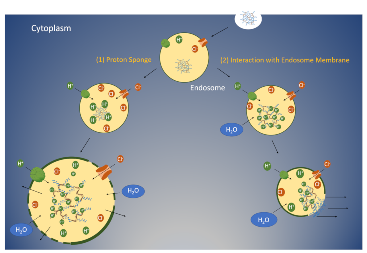
The archetypes here are cationic polymers such as PEI, along with histidine-rich peptides and LAH4-type peptides. The appeal is that the mechanism is intuitive and the design lever is clear: raise buffering capacity within the endosomal acidification window and destabilize the membrane.
This route is contested more sharply than the debate framing suggests. Direct measurements of endosomal pH and volume have repeatedly failed to reproduce the osmotic swelling the model predicts, and several groups have argued that proton-sponge polymers escape by other means — or that buffering capacity and escape efficiency simply do not correlate (Benjaminsen et al., Mol Ther 2013; Vermeulen et al., ACS Nano 2018). Endosomal ion flux and membrane repair in living cells are also far more active than the model assumes. The honest position is that the proton sponge remains a useful design heuristic whose mechanistic status is unresolved. Separately, membrane disruption strong enough to be effective is often strong enough to be cytotoxic.
For a chronic disease requiring long-term dosing this is not an attractive development path. Newer EEPs therefore pursue not stronger buffering, but a more finely tuned pH-response and membrane-perturbation window.
Pore formation and membrane disruption: membrane-active peptides in acidic endosomes
The second mechanism is pH-triggered membrane insertion, pore formation, and membrane perturbation. Many peptides are random-coil or relatively inert at neutral pH but undergo conformational change on reaching the acidic endosome, forming an amphipathic α-helix. A hydrophobic face and a hydrophilic face allow insertion into the bilayer, producing local thinning, pore formation or increased permeability, through which nucleic acid cargo escapes to the cytosol.
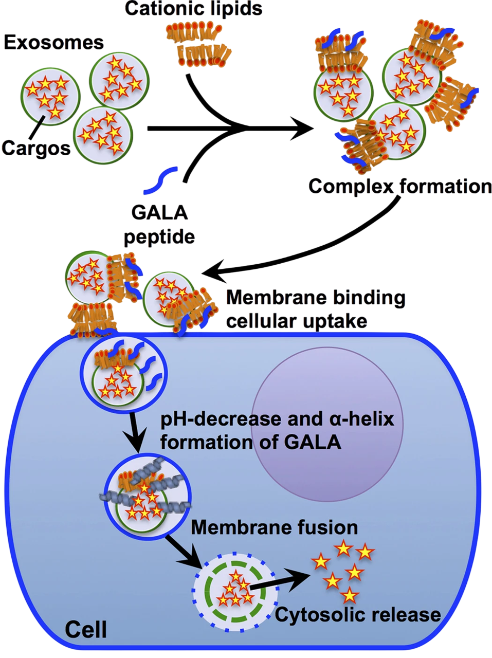
This route borrows explicitly from viral escape. The HA2 fusion peptide of influenza haemagglutinin undergoes conformational rearrangement at low pH and promotes fusion of the viral and endosomal membranes; Research peptides such as GALA and INF7 exploit the same logic. The core advantage is that the peptide stays inactive in blood and the extracellular space and is activated only after endosomal acidification. Tuning amino acid composition, hydrophobicity, helical propensity and pKa couples membrane activity to the acidification process and limits systemic toxicity.
The development difficulty is symmetric: too little membrane activity and escape is insufficient; too much and the plasma membrane or mitochondrial membranes are damaged. A developable EEP must find the window between "escapes enough" and "stays gentle enough". This is why many academically potent membrane-active peptides never translated — impressive in cell culture, but on systemic dosing they meet plasma stability, complement activation, renal exposure and tissue toxicity.
Cyclic peptides and membrane domain remodeling: Entrada's EEV model
Entrada has proposed a distinct mechanistic model for EEV™, described principally in company conference presentations: after the cyclic CPP binds the endosomal membrane, CPP-rich lipid domains form; These induce vesicle-like structures to bud from the membrane and subsequently collapse into amorphous peptide–lipid aggregates, releasing cargo. The acidic environment is proposed to promote this by increasing CPP–membrane binding affinity. It should be read as a sponsor-proposed working model: the supporting data are largely from company decks and model membrane systems, and it has not been independently reproduced in peer-reviewed literature.
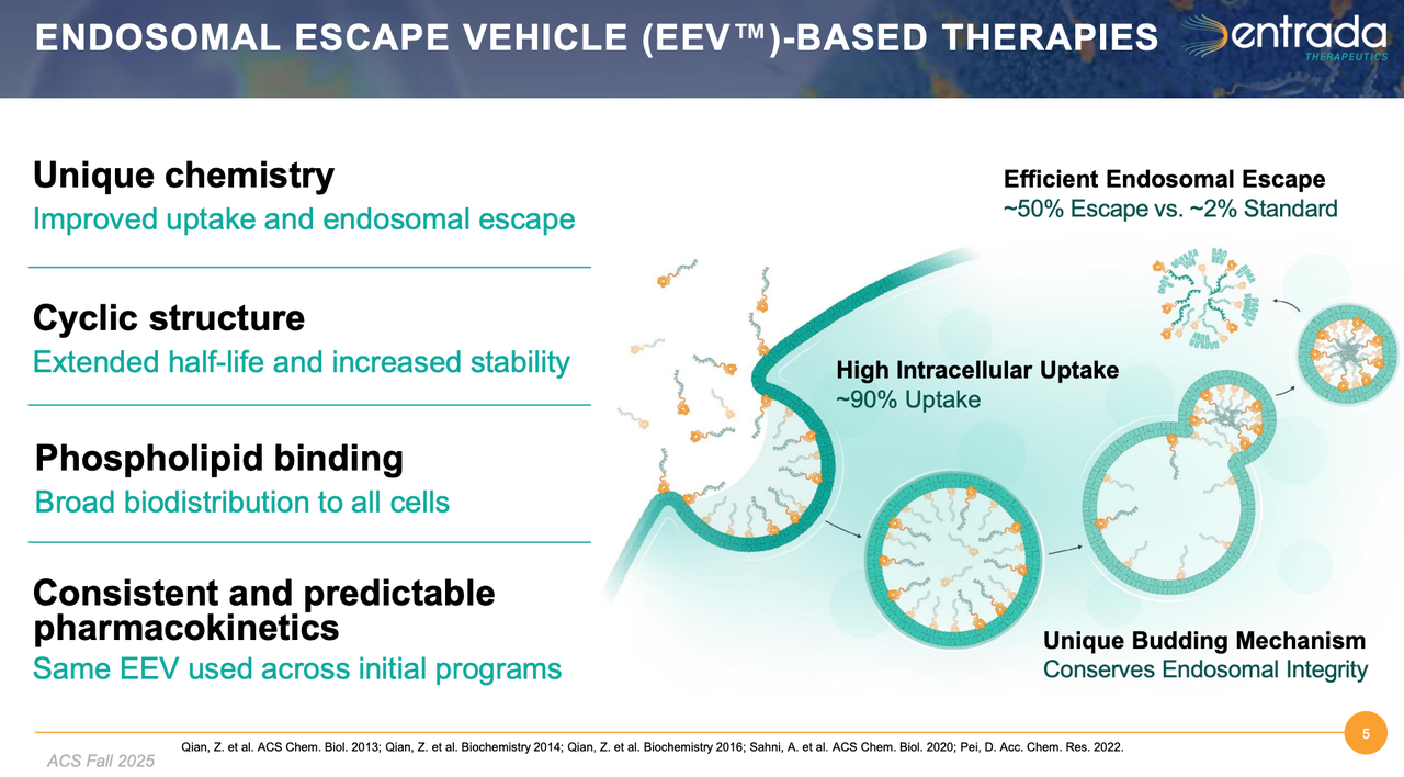
This differs from a proton sponge or a simple pore: it is organized interaction between cyclic peptide and membrane lipid inducing local membrane remodeling. If it holds, the implication is significant, because release would not require large-scale endosomal destruction and could reconcile efficiency with safety.
The defensible statement at this stage is that EEV represents a class of cyclic-peptide escape strategy distinct from conventional linear CPPs, whose mechanism may involve endosomal membrane binding, domain formation and local remodeling (vesicle budding and collapse, VBC) rather than proton sponge effects or large-scale pore formation alone.
Mechanistically, the goal of an EEP is not to destroy the endosome. The ideal is a brief, gentle release window opened in the right cell, at the right time, in the right acidic environment. Too little escape and there is no drug effect; too much and safety becomes the bottleneck. That is the hardest part of building an EEP platform.
4. How PepGen and Entrada are Advancing EEPs
PepGen and Entrada have both taken EEPs into human studies. PepGen follows a linear engineered CPP route (platform: EDO); Entrada follows a cyclic CPP route (platform: EEV). Both use PMO as the primary payload and both focus on neuromuscular disease. Both set out to show that optimizing endosomal escape and intracellular delivery could substantially raise the efficacy ceiling of conventional PMO — a proposition that has now been tested in DMD and only partly supported.
PepGen EDO: engineering the linear CPP approach
PepGen's core platform is EDO (Enhanced Delivery Oligonucleotide) — in essence a PMO conjugated to an optimized cell-penetrating peptide, forming a peptide–oligonucleotide conjugate. Relative to naked PMO, EDO aims to substantially improve tissue penetration, myocyte uptake, endosomal escape, and nuclear delivery.
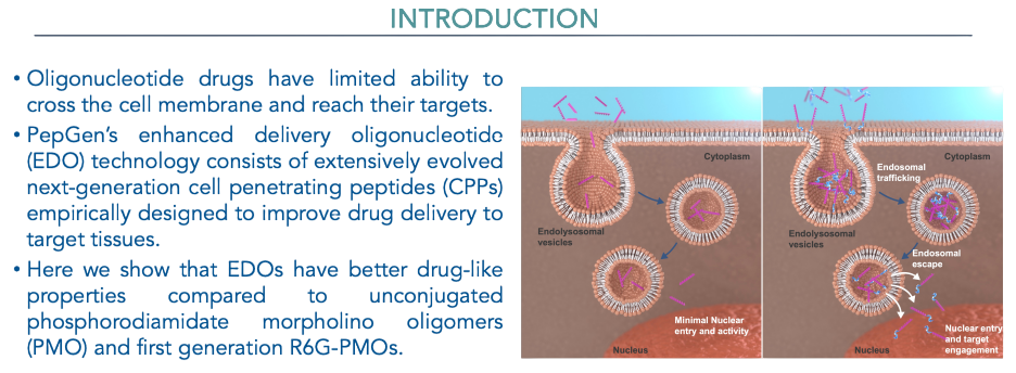
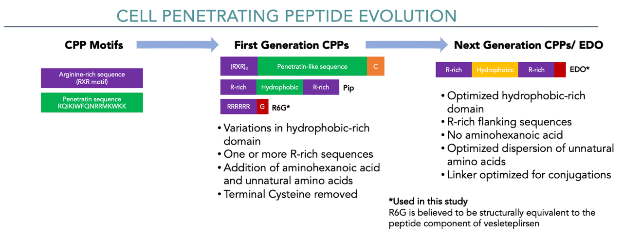
PepGen's peptide design is not simply more arginine. According to company materials, the EDO peptide is typically a short (<17 amino acid) linear engineered CPP containing an optimized poly-Arg region (roughly 5–6 arginine flanking sequences), unnatural amino acids, and a hydrophobic core that assists endosomal escape.
Compared with first-generation CPPs (which used RXR motifs, amino hexanoic acid spacers and similar architecture), the design goal for next-generation EDO peptides is to retain efficient uptake while reducing the toxicity and instability of conventional highly cationic CPPs, so as to achieve clinically viable tolerability.
Mechanistically, PepGen has done systematic work. The company has used cellular imaging to track how EDO colocalization with endosomal and lysosomal markers such as Rab5, Rab7 and LAMP1 changes, as a read on whether EDO is effectively released from the endocytic pathway. It has also used a HiBiT split-luciferase system to quantify cytosolic delivery. Published posters show that next-generation EDO improves on first-generation CPP–PMO in uptake, endosomal escape and functional exon skipping.
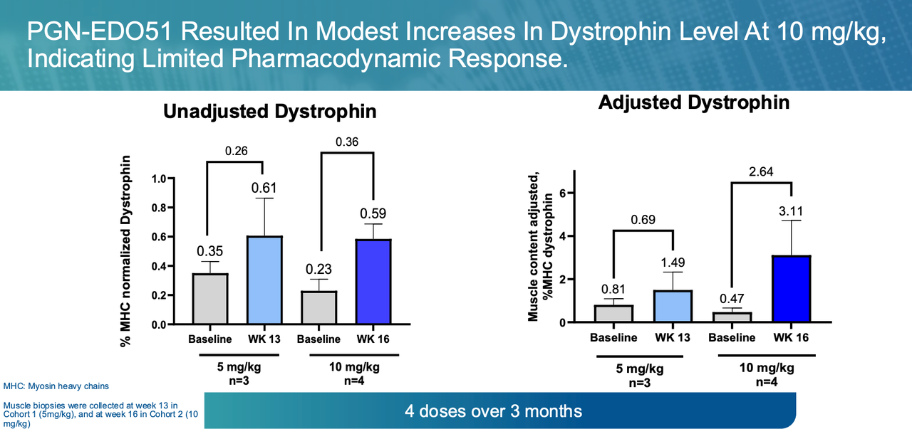
In DMD, PepGen developed PGN-EDO51 for exon 51 skipping. In May 2025, the company announced that in the 10 mg/kg cohort of CONNECT1 (n=4), PGN-EDO51 raised exon-51-skipped transcripts to 4.26% (a mean increase of 3.5%) but total dystrophin reached only 0.59% of normal (a mean increase of 0.36%). PepGen judged this insufficient, voluntarily discontinued PGN-EDO51, stopped dosing in CONNECT1, closed CONNECT2 without dosing any participants, and wound down all DMD-related R&D. The setback is a clean illustration of the field's central translational risk: transcript-level exon skipping and protein-level dystrophin restoration are not linearly related. A 3.5-percentage-point gain in skipped transcript yielded 0.36 percentage points of dystrophin — roughly a tenfold loss between the RNA and protein readouts. Any platform arguing from a transcript biomarker to clinical benefit inherits this gap, including the DM1 splicing data discussed below.
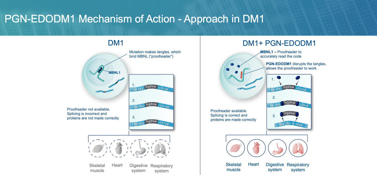
In DM1, PGN-EDODM1 better demonstrates the platform value of EDO. DM1 is caused by expanded CUG repeats in the DMPK transcript forming a toxic RNA structure that sequesters splicing factors such as MBNL1, producing widespread mis-splicing. PGN-EDODM1 uses a PMO to bind the pathogenic DMPK RNA and release the splicing factors, correcting downstream mis-splicing. This mechanism places unusually heavy demands on nuclear delivery: the drug must not only enter the myocyte, but also escape the endosome and reach the nucleus to engage the abnormal RNA. Thus, dose-dependent splicing correction in patients indicates strong evidence of EDO delivery.
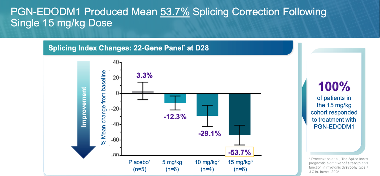
In FREEDOM-DM1 (Phase 1 SAD), PGN-EDODM1 showed dose-dependent splicing correction at day 28 on a 22-gene panel: 12.3% at 5 mg/kg (n=6), 29.1% at 10 mg/kg (n=4) and 53.7% at 15 mg/kg (n=6). PepGen describes the 15 mg/kg result as higher than any previously reported splicing correction in DM1. These are small cohorts within a randomized placebo-controlled SAD study, and one participant was excluded from analysis for a splicing index outside the pre-specified assay range at both baseline and day 28.
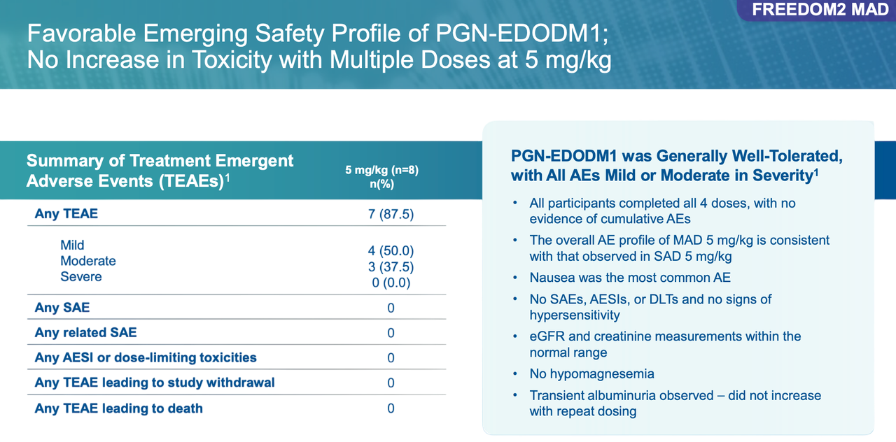
In FREEDOM2-DM1 (Phase 2 MAD), the lowest 5 mg/kg cohort showed mean splicing correction of 7.3% (n=6) against 6.8% on placebo (n=2) — essentially no separation from placebo at this dose. A post-hoc analysis excluding one notable splicing outlier gave 22.9% (n=5). Safety and tolerability were favorable, with all AEs mild or moderate, no SAEs and no evidence of cumulative toxicity on repeat dosing. On that basis the company advanced to the 10 mg/kg cohort, with data guided to 2H 2026.
On safety, PepGen's route carries risks that require active management. In FREEDOM-DM1 Phase 1, the 15 mg/kg cohort showed transient (~48 hours), asymptomatic changes in renal biomarkers, eGFR decline, creatinine rise, proteinuria, which met dose-limiting toxicity criteria in one participant; the events were mild-to-moderate and resolved without intervention. In FREEDOM2-DM1, the 5 mg/kg MAD cohort showed all AEs mild or moderate, no SAEs and no cumulative toxicity on repeat dosing. This history should be read alongside the DMD program: CONNECT2 was placed on FDA clinical hold following safety findings before CONNECT1 read out, and the renal signal is the recurring liability of highly cationic peptide–oligonucleotide conjugates rather than an incidental finding. A safety window has been demonstrated at 5 mg/kg on repeat dosing, and the renal margin at higher repeat doses is precisely what the 10 mg/kg cohort will test.
Entrada EEV: engineering the cyclic CPP approach
Unlike PepGen's linear CPP route, Entrada represents a different EEP approach: cyclic cell-penetrating peptides. The platform is named EEV™ (Endosomal Escape Vehicle), reflecting the company's focus on endosomal escape as the core problem from the outset, rather than treating the delivery peptide as an accessory modification to the oligonucleotide. EEV is built on cyclic CPPs, which offer greater conformational stability, better protease resistance, more controllable membrane interaction, and better tractability for systematic medicinal chemistry optimization. The specific escape mechanism proposed for EEV — CPP-rich domain formation, vesicle budding and collapse — is discussed in §3.3 above.
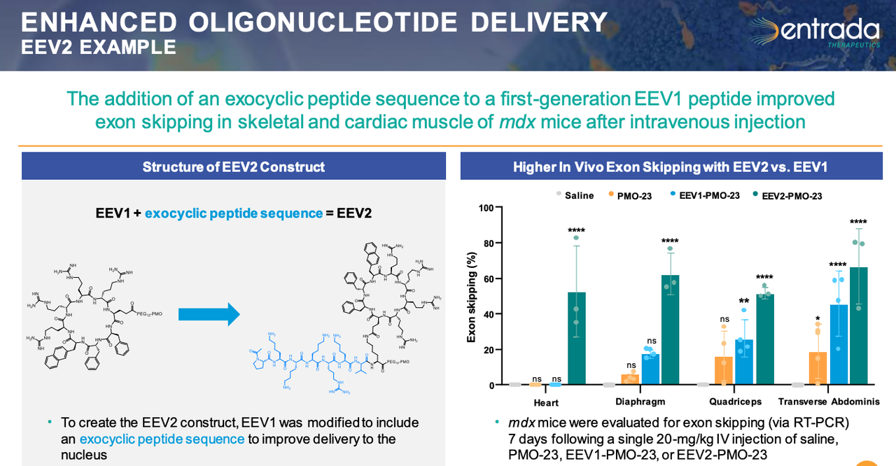
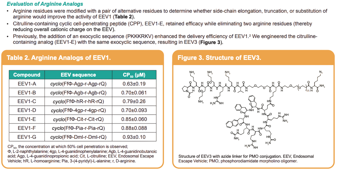
According to Entrada's public materials, the EEV platform has gone through several generations. EEV1 already enhanced PMO cellular uptake and functional delivery. An exocyclic peptide was then added, including an SV40-derived nuclear localization sequence (NLS, PKKKRKV), to produce EEV2 and improve nuclear delivery; company data report roughly a 3.3-fold increase in nuclear localization and a 2.6-fold increase in total cellular uptake in C2C12 myotubes relative to EEV1. EEV3 then substituted some cationic residues in the macrocycle (for example introducing neutral residues such as citrulline) to maintain delivery while lowering toxicity and improving pharmacokinetics and tissue distribution.
This iteration pattern is worth noting: EEP platform development is not a matter of maximizing positive charge, but of using medicinal chemistry to balance membrane affinity, escape efficiency, tissue exposure, plasma stability, renal exposure and immunological safety.
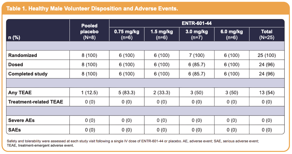
Entrada's lead program is ENTR-601-44 for DMD exon 44 skipping, an EEV–PMO conjugate designed to deliver an exon 44 skipping PMO more efficiently into skeletal and cardiac muscle. In preclinical models it produced exon skipping and dystrophin restoration signals in both skeletal muscle and heart. Relative to naked PMO, the central claim for EEV–PMO is broader muscle tissue coverage, particularly the potential for cardiac delivery.
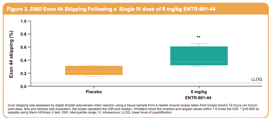
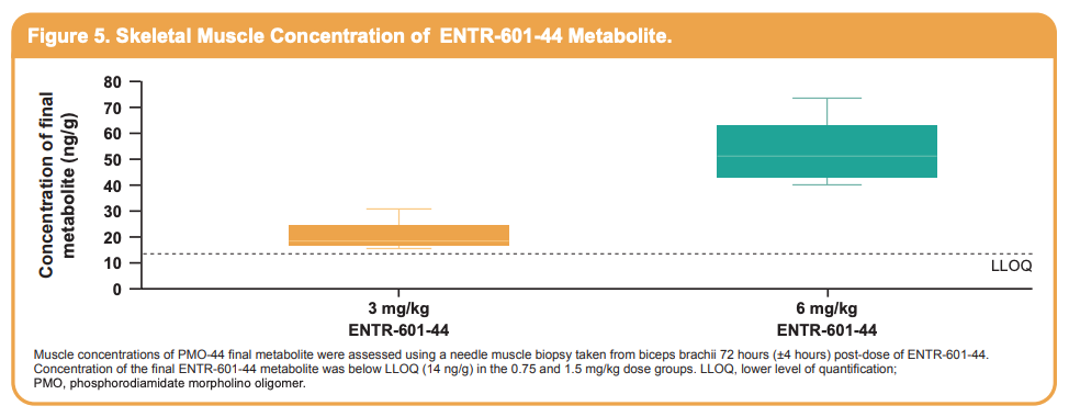
In humans, ENTR-601-44 was generally well tolerated in the ENTR-601-44-101 healthy male volunteer SAD study (32 subjects across four cohorts: 0.75, 1.5, 3 and 6 mg/kg; 6 active and 2 placebo per cohort), with no serious adverse events, no drug-related adverse events, and no clinically relevant changes in renal toxicity biomarkers at the highest dose. In the 6 mg/kg cohort, the final PMO metabolite was detected in all six subjects (mean 53.8 ng/g, range 40–73.5 ng/g) and mean exon 44 skipping was 0.44% (range 0.3–0.65%), which was statistically significant versus placebo (p<0.005, Mann–Whitney U). The absolute values are low, but the result establishes an on-target molecular effect of EEV–PMO in human muscles.
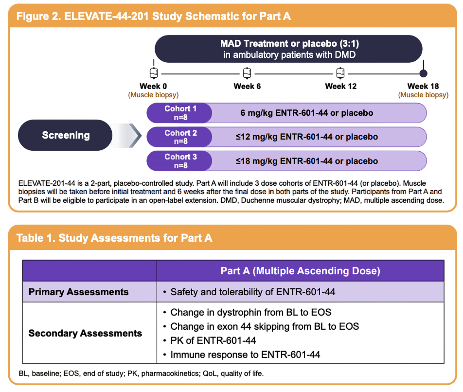
ENTR-601-44 was placed on FDA clinical hold in December 2022; The hold was removed in February 2025, alongside MHRA authorization in the UK for ELEVATE-44-201. Cohort 1 of ELEVATE-44-201 has since read out (7 May 2026): ambulatory participants aged 6–17, randomized 3:1 to three doses of 6 mg/kg or placebo, showed a 2.36% increase in dystrophin from a 4.00% baseline and a 2.31% increase in exon skipping from a 2.66% baseline, together with a statistically significant improvement in time-to-rise velocity versus placebo (p<0.05). Plasma exposure in this pediatric cohort was lower than in healthy adults, prompting revised PK modelling and escalation to 12 mg/kg in Cohort 2, with further data guided to end-2026.
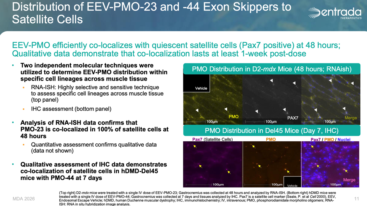
Entrada has another potential differentiator: in preclinical models, EEV–PMO appears to reach Pax7⁺ muscle satellite cells, with PMO colocalization still detectable after extended periods. The rationale is that DMD and DM1 involve not only mature myofibers but ongoing regeneration, repair and progenitor pool exhaustion, so a delivery system that reaches satellite cells might influence long-term disease course rather than only short-term splicing or protein expression in mature fibers. Entrada has begun invoking satellite cell access in 2026 as an explanation for why modest dystrophin gains in ELEVATE-44-201 Cohort 1 were sufficient to improve time-to-rise velocity. That inference is currently supported by preclinical colocalization data plus a functional signal from a small cohort, with no direct human evidence of satellite cell engagement — it remains a differentiating hypothesis, not a demonstrated mechanism.
At an industry level, Entrada has won strategic validation from large pharma. Vertex and Entrada announced a collaboration in December 2022, which became effective in 2023 after regulatory clearance; it was center on DM1 (ENTR-701 / VX-670) with options on additional targets rather than being a platform-wide grant. The read-through is still that large pharma is attending to intracellular delivery as a bottleneck, but the deal should be described for what it is: indication-anchored, with platform optionality, not a blanket endorsement of EEV across muscle disease.
If PepGen's EDO route represents clinical testing of linear engineered CPPs, Entrada's EEV represents an attempt to turn cyclic peptide delivery into an iterable, extensible technology platform. Together they constitute the two most important technical routes in current EEP clinical development.
Beyond PepGen and Entrada: the broader landscape of endosomal escape technologies
PepGen and Entrada are the most representative clinical-stage companies, but endosomal escape is being worked on far more widely.
Peptirna's approach is to let cationic peptides self-assemble with anionic siRNA into nanoparticles, attempting to solve siRNA encapsulation, cellular uptake, endosomal escape and extrahepatic delivery simultaneously.
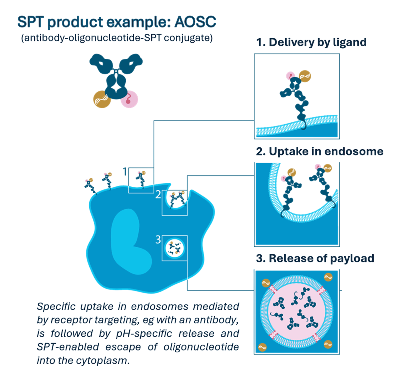
Sapreme Technologies takes a non-peptide route: its SPT platform uses saponin molecules to promote endosomal escape. Saponins are amphipathic glycosides that interact with membrane cholesterol in the acidic endosomal environment, altering membrane permeability and releasing contents. This is preclinical, but it makes the general point: endosomal escape is not a peptide-specific problem. Any nucleic acid drug that depends on endocytosis to enter cells must solve it.
LNPs are themselves an efficient endosomal escape system. Ionizable lipids protonate in the acidic endosome, acquire positive charge, and interact electrostatically with anionic lipids in the endosomal membrane, forming non-bilayer structures (such as the inverted hexagonal H₁₁ phase) that drive fusion and destabilization and release mRNA or siRNA into the cytosol. Here the escape module comes from the lipid, not a peptide — though it is worth noting that quantitative estimates of LNP escape efficiency are also low, in the low single-digit percent range.
Ionis and other ASO companies have long studied the intracellular trafficking and endosomal release of PS-ASOs. A PS-ASO carries no external peptide or saponin escape module, but its phosphorothioate backbone increases protein binding — to cell surface proteins, endosomal proteins and membrane structures — so that after entering cells through multiple endocytic routes, a small fraction undergoes productive release from late endosomes. Work from Ionis indicates that LBPA/Alix-mediated back-fusion of intraluminal vesicles, COPII/STX5-related transport, and M6PR/GCC2-mediated Golgi–endosome trafficking all influence the efficiency of PS-ASO release from late endosomes. Overall efficiency is very low, but for an RNase H ASO, even a very small fraction reaching the cytosol or nucleus may be enough to support clinical activity.
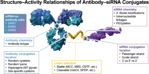
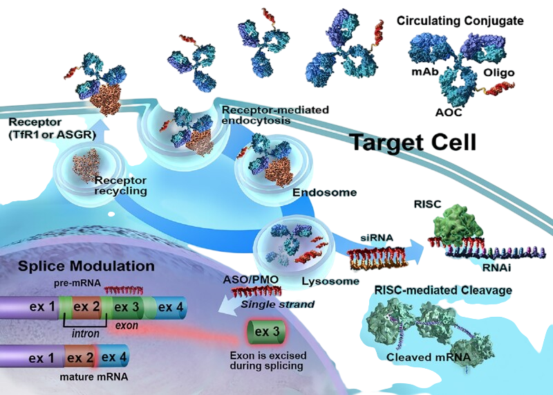
Avidity Biosciences' AOC platform represents another non-peptide targeted delivery route. AOC conjugates a monoclonal antibody to siRNA or PMO, using antibody recognition of TfR1 on muscle cells to improve oligonucleotide entry into skeletal and cardiac muscle via receptor-mediated endocytosis. Unlike EEPs or saponin-type escape enhancers, Avidity's public materials emphasize targeted entry into the intended tissue and cell. Oligonucleotide that has entered the endosome still has to be released, and the specific escape mechanism has not been extensively disclosed by the company.
Taken together, these routes show that endosomal escape is a shared bottleneck across the nucleic acid delivery field. The advantage of the EEP is that it can be engineered at the level of amino acid sequence and medicinal chemistry, which suits it particularly well to forming structurally defined conjugates with PMOs and ASOs.
5. Defining the Moat, Limits, and Future of EEPs
The patent moat around an EEP platform looks like peptide sequence but is in substance the whole delivery chemistry.
The first layer is sequence and conformational protection. PepGen's position centers on engineered linear CPPs: charge distribution, hydrophobic core, unnatural amino acid combinations and specific sequences. Entrada emphasizes cyclic CPPs, exocyclic peptides, nuclear localization sequences and citrulline substitution.
The second layer is conjugation chemistry and CMC. Where the peptide attaches to the PMO, ASO or siRNA, and how the linker is designed, directly affects drug properties and product homogeneity. Peptide–oligonucleotide conjugates are smaller than antibodies but still raise conjugation site, purification, stability and scale-up problems.
The third layer is combination protection across indication and regimen. Different indications correspond to different nucleic acid sequences, and the combination of delivery peptide with a specific cargo and a specific disease, together with IV dosing frequency, dose range and tissue distribution characteristics, can create new scope of protection.
So EEP competition is not about who found a peptide that crosses membranes; first-generation CPPs have long been public knowledge. The real moat is who can close the loop across peptide sequence, conjugation chemistry, tissue distribution, safety window and human efficacy.
Endosomal escape is also not a "more is better" parameter. In chronic diseases such as DMD and DM1 requiring long-term repeat dosing, the safety window matters more than single-dose potency. More arginine or lysine does increase uptake, but also increases plasma protein binding, ECM retention, renal tubular injury and non-specific tissue accumulation. PepGen has optimized the Arg domain and introduced unnatural amino acids and a hydrophobic core to improve stability; Entrada has used cyclisation, exocyclic peptides and neutral residue substitution to tune membrane interaction. The direction is the same in both cases, not maximal membrane disruption, but opening a release window brief and gentle enough to be tolerable.
On current evidence, the best-fitting application for EEPs is oligonucleotide delivery in neuromuscular disease, for three reasons. First, DMD and DM1 involve skeletal muscle and myocardium throughout the body and require broad tissue coverage. Second, myofibers are large and the extracellular matrix is dense, so macromolecular delivery shows marked diffusional heterogeneity; peptide–oligonucleotide conjugates are far smaller than antibodies and should, in principle, penetrate deep tissue better. Third, these indications have clear pharmacodynamic readouts — exon skipping, dystrophin restoration and motor function in DMD; splicing correction and myotonia in DM1 — which makes it possible to test whether a delivery platform works.
EEPs may extend to heart, lung, tumor and CNS, each with its own obstacles. Cardiac safety requirements are extremely high; the lung presents a mucus barrier and immune reactivity; tumors pose a tension between penetration and non-specific toxicity; The CNS requires solving BBB penetration first. The future of the EEP is therefore not to become a universal delivery platform but to act as a key module inside different delivery systems, combined with POC, LNP, AOC and even gene editing tools.
6. Key Takeaways
Nucleic acid delivery is entering a new phase of competition.
The first phase was organ access. GalNAc established the liver as a commercially validated destination for oligonucleotide therapeutics. The second was extrahepatic reach, with AOC, POC and LNP platforms extending delivery toward muscle, heart, and the CNS. The next is intracellular accessibility: reaching the right tissue is no longer enough. The payload must enter the right cells, escape the endosome, and reach the cytosol or nucleus in a pharmacologically active form.
EEPs sit directly at this transition.
Their value comes from where they act — at one of the most consequential, yet historically underestimated, steps in the delivery cascade. Cellular uptake alone does not equal productive delivery. A payload trapped in the endosomal system may generate an impressive microscopy signal, but little pharmacology. Only after release into the cytosol or nucleus can a nucleic acid therapeutic engage its target and translate exposure into biological effect.
PepGen and Entrada are turning endosomal escape from a delivery concept into a clinically tested platform strategy.
PepGen and Entrada illustrate two distinct approaches to solving this problem: engineered linear CPPs through EDO and cyclic CPPs through EEV. Their clinical programs also highlight an important reality: improving intracellular delivery is necessary, but not sufficient. PepGen has demonstrated dose-dependent splicing correction in DM1, while its DMD experience showed that exon skipping does not automatically translate into meaningful dystrophin restoration. Entrada, meanwhile, has reported target engagement in human muscle and early evidence of dystrophin production in patients.
The lesson is broader than either platform.
Endosomal escape may raise the ceiling of nucleic acid delivery, but clinical success still depends on converting molecular engagement into protein-level and ultimately functional benefit. That transcript-to-protein transition is where delivery biology, target biology, and therapeutic design converge.
The strongest delivery platforms of the future are unlikely to win through a single carrier concept. They will integrate tissue targeting, tissue penetration, cellular uptake, endosomal escape, subcellular localization and safety into one coordinated system.
The endpoint of delivery is not the plasma membrane.
It is the compartment where the drug can actually work — and endosomal escape is becoming one of the most important gates on the way there.

Our mission is to democratize computational drug discovery tools and empower researchers and organizations worldwide.